Portable and easy-to-use manikin for teaching and learning heart, breath, and bowel sounds. Although used in many simulation centers, the manikin is also easily moved into a classroom or auditorium for group instruction. The computer software interface is easily projected into any smart classroom. The software includes phonocardiograms, correct anatomical locations, and written lessons for each sound. Features include: • Carotid pulse • Customized volume adjustments • Large library of sounds includes 35 heart, 21 breath, 16 bowel, and four carotid bruit sounds, all heard at the correct anatomical location (both anterior and posterior), along with 16 heart/breath combination sounds • Pediatric sounds – includes ASD, PDA, VSD, pulmonary hypertension, pulmonary stenosis, and Tetralogy of Fallot • Phonocardiogram • Portability – lightweight manikin weighs only 23 lbs. • Preprogram your own password-protected lectures and scenarios • Smart classroom/auditorium ready • Students use their own stethoscope • Variable breath sounds • Variable heart and respiration rates Includes male manikin, Dell laptop computer with SAM II software installed, 196-page lesson guide, and operator’s manual. Shipped directly from our supplier. Allow extra delivery time.
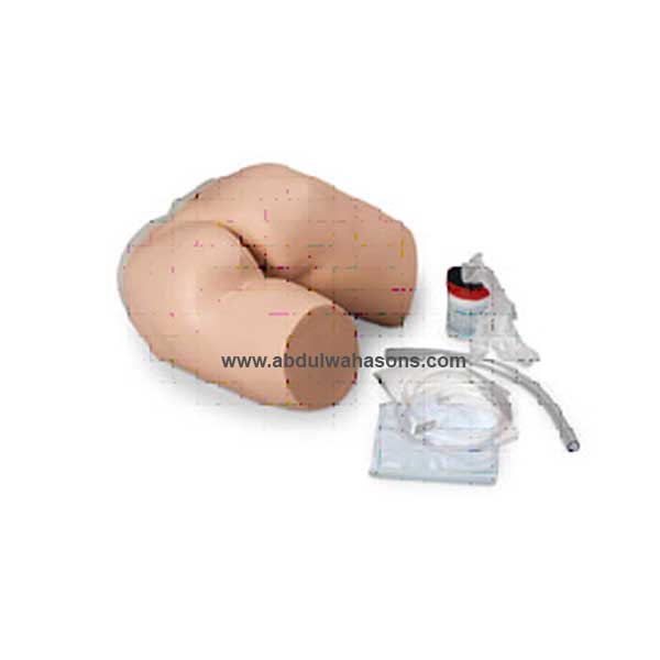
Manikin has all the same features as the complete CRiSis™ Manikin (LF03953U with the addition of auscultation sites with heart and lung sounds, plus an ECG Simulator LF03670U). For auscultation training, the instructor selects from a menu of heart and lung conditions by wireless remote control. The manikin presents itself as a real patient without visible auscultation sites. The student must palpate to identify correct auscultation locations, and will hear different heart and lung sounds as the SmartScope™ is moved to different locations on the manikin. A diagnosis of the condition selected by the instructor can be made by comparing the variations in sounds occurring at different sites – just like a real patient! Lung sounds can be detected at five anterior and two midaxillary locations and students can practice auscultation at six anterior heart sites. The remote control does not have to be pointed directly at the manikin or SmartScope™ to operate. One remote control will operate multiple sets of SmartScopes™ and manikins simultaneously. Great for group instruction. Listen to the sounds by using either the single or dual user headpieces on SmartScope™ or by connecting to an amplified speaker (not included, see SB20146U). Range on the unit is up to 100 ft. of remote access. Adult CRiSis™ Auscultation Manikin includes Complete CRiSis™ Manikin with auscultation sites, one remote control with LCD display, one SmartScope™ with single- and dual-user headpieces, hard storage case, and ECG Simulator (LF03670U). Requires two “AA” and two “AAA” batteries (included).
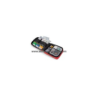
This new and improved AED trainer with plug-in module has all the functions needed to teach students about AED operation – without breaking your equipment budget. This lightweight, value-priced trainer offers bilingual scenarios (English and Spanish), a fully functional remote control, volume adjustment and pause functions, adult and child connectors, and pads. Includes eight pre-programmed scenarios to simulate various cardiac arrest cases. Audible metronome paces CPR chest compressions. Audio port allows you to connect speakers so the entire class can hear scenarios as they play out – extending training even while they wait. Plug-in design allows for easy upgrades and language changes. Comes in a 10-1/2″ x 8″ x 2-3/4″ nylon carrying case. Base requires three “AA” batteries (not included). Remote control requires two “AAA” batteries (not included). AED trainer measures 7″ x 5-3/4″ x 2″. Meets 2005 ECC guidelines. against defects.
A life-size human torso with interchangeable male and female genital organs with embryo in third month of pregnancy. Dissectible into 24 parts: head, half brain with arteries, eye with optic nerve, female breast cover, two lung halves, heart (2 parts), stomach (2 parts), liver with gallbladder, large intestine with appendix flap (3 parts), small intestine, kidney half, female genital organs (2 parts) with third month embryo, male genital organs (4 parts), and torso. Unbreakable plastic. 15″ x 10″ x 34″ mounted on base. Torso teaching guide included
Abdul Wahab & Sons provides advanced scientific and medical education solutions, including VR/AR, holograms, 3D Anatomy Tables, and PowerLab systems. AWS serve hospitals, research labs, medical schools, and universities with precision instruments, skill labs, and immersive learning tools empowering the future of healthcare and science.


Reviews
There are no reviews yet.