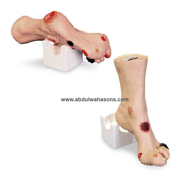
Wound Foot
Product Description
Molded from a real foot for a true-life experience when assessing the various wounds. Twenty different conditions are presented so you can see and understand how they are different. Great care has been taken to color each wound just as you would see it on a patient. Once the different etiologies are understood, you can discuss and devise treatment plans that will deliver optimized patient care. The following wounds and abnormalities are included: pressure ulcers, Deep Tissue Injury, callus, amputated toe, gangrene, maceration, partial thickness wounds, corn, ingrown toenail, blister, hammer toes, and skin stapled wound. Measures 12-1/2″ x 8″ x 3-5/8″. Comes with positioning base for “hands free” access to all the sites when applying dressings or teaching.
Related products
-
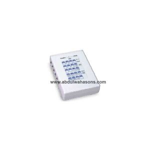
Code Simulator 12-Lead Arrhythmia Simulator – ZOLL
Pace and defibrillate directly into this interactive 12-lead ECG simulator through your hands-free defibrillation cable. Connects directly to monitor • Does not work with manikin • Simulate electrical capture with your external pacer. Press simulator capture key to select one of four preset pacing capture levels: 70, 80, 90, or 100mA. When pacer current is greater than selected capture level, paced beats appear on your monitor. Waveforms for pacing include: Sinus Brady (two), 1st degree A-V block, 2nd degree type I A-V block, 2nd degree type II A-V block, 2nd degree type II A-V block with PVCs, and 3rd degree A-V block. • Simulate cardioversion with your manual, semi-automatic, or automatic defibrillator. Activate “convert” feature to select - before defib discharge - another rhythm to run immediately after defib discharge. Waveforms for defibrillator training include: V. Fib., V. Tach (high rate), V. Tach (low rate), Torsade, A. Fib, A. Flutter, PSVT, Sinus Tach, Sinus Rhythm, Sinus Rhythm with PVCs, Asystole, and NSR. • Generate realistic 12-lead ECG rhythms. Connect your ECG cable to simulator’s 10 ECG snaps. Independent chest lead and limb lead ECG signals create realistic 12-lead ECGs for each rhythm. Generate ST segment and T wave abnormalities including: Anterior MI, Inferior MI, Antero-Septal ST Elevation, Anterior ST Depression, Lateral ST Elevation, and Inferior ST Elevation. • LED indicators: pacer pulse detection, defib discharge detection, and low battery.
-
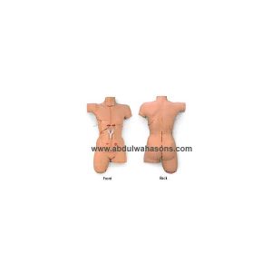
“Surgicall sally” Bandaging Simulator
A readily available patient with 14 surgical wounds including a Mid-Sternal Split with two simulated drains, a sacral Decubitus Ulcer-stag 2, and a leg Amputation stump. The skin has also been reformulated so it is drier, allowing the bandages to adhere better. Wound closures feature surgical staples for the Mid-Sternal Split, Tharacotomy, Nephrectomy, Laparoscopy, Abdominal Hysterectomy, and Amputation stump. Staples and sutures cannot be removed.
Ideal for teaching wound management skills, dressing change, and bandaging techniques. This simulator is just like a real patient, except the simulator remains motionless, never complains, and features wounds that won’t bleed and closures that won’t rip out. Students can conveniently practice wound management techniques, cleaning, and dressing change.
Surgical Sally is a female replica and measures 29” L*17”*8”D.
Special formulations have been used to create the most realistic responds look and feel possible. The flexible, flesh-colored skin realistically responds with adhesive and all types of bandaging procedures. Comes complete with hard carry case. Size: 33” *25” *12”. wt.24lbs.
-
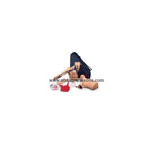
Peter PICC Line
Product Description
The only available teaching model which permits the practice of placing Peripherally Inserted Central Catheter Lines (PICC Lines). The rapid acceptance and use of PICC Lines creates a need to be able to properly teach this procedure. Peter PICC Line™ allows both the teaching and assessment of this insertion technique. This portable teaching model has translucent arm skin that allows the visualization of the anatomically correct cephalic, basilic, and median basilic veins. The axillary jugular, subclavian, and superior vena cava veins are also present, plus a movable chin simulates the occlusion of the jugular to prevent the PICC Line from taking this path. Palpable ribs permit practice in measuring proper catheter length to the correct intercostal space. The model also permits the measurement of catheter length outside of the body and the confirmation of proper placement by viewing the distal tip of the catheter in the viewable superior vena cava. Replaceable, barely translucent arm skin allows visualization of underlying veins. Includes soft carrying case. A great tool for training and skill assessment. Blue veins are made of natural latex. Measures 17″ x 22″ x 5-1.2″.


Reviews
There are no reviews yet.